Why Do Fluids Leave The Capillaries At The Arterial End
A clear, physiology-first explanation of why fluids leave the capillaries at the arterial end, with pressure math, clinical clues, and safe next checks.
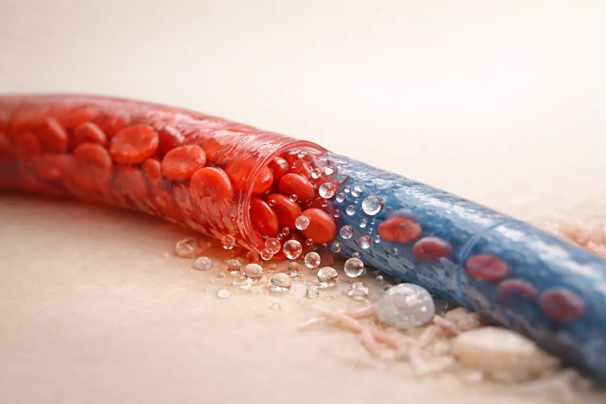
Quick answer: Starling forces make fluid exit at the arterial end
Fluids leave capillaries at the arterial end because capillary hydrostatic pressure there is higher than the opposing plasma (oncotic) pressure, creating a net outward force that pushes plasma water into the interstitial space. In simple terms: the push from blood pressure on the arterial side overcomes the pull from blood proteins, so filtration dominates at the upstream portion of the capillary bed.
Physiologists summarize this balance with Starling forces: capillary hydrostatic pressure (Pc) versus capillary oncotic pressure (πc), plus smaller contributions from interstitial hydrostatic (Pi) and interstitial oncotic pressure (πi). At the arterial end Pc is large enough that NFP (net filtration pressure) is positive, so fluid exits the capillary.
This outward flow is normal and necessary: it delivers dissolved nutrients and oxygen into tissues. Most of the filtered fluid is recovered later at the venous end or returned to circulation via the lymphatic system. The immediate takeaway: higher hydrostatic pressure at the arterial end causes outward movement of fluid.
- Positive net filtration pressure (NFP) at the arterial end drives fluid out.
- Hydrostatic pressure (blood pressure in capillaries) is higher on the arterial side.
- Plasma proteins (oncotic pressure) oppose filtration but are relatively constant along the capillary.
- Normal balance: filtration at arterial end, reabsorption at venous end, lymphatic return of excess fluid.
What it means
Saying that fluids leave the capillaries at the arterial end describes a dynamic balance in every capillary bed. Capillaries are thin-walled vessels designed for exchange; fluid movement across their walls helps deliver oxygen, glucose, and hormones while removing metabolic byproducts. The fact that fluid leaves at the arterial end means tissues get a steady supply of plasma water and dissolved materials where perfusion begins.
Reabsorption and lymphatic drainage complete the cycle. Toward the venous end of the same capillary the hydrostatic pressure falls (due to resistance and fluid loss) and oncotic pressure stays roughly the same, so the net force shifts toward inward movement and fluid returns to the vascular compartment. Any excess that is not reabsorbed is collected by lymphatic capillaries and returned to the circulation.
In practical terms, this mechanism keeps interstitial spaces moist but not flooded. If the balance is shifted—by high capillary pressure, low plasma proteins, increased capillary permeability, or blocked lymphatics—filtration can overwhelm clearance and cause edema. So the arterial-end filtration is physiologic, but it can be amplified by disease or injury.
- Arterial-end filtration supplies tissue with plasma water and solutes for exchange.
- Venous-end reabsorption and lymphatics recover filtered fluid to prevent swelling.
- Disturbances in pressure, oncotic forces, permeability, or lymph flow produce edema.
- Exchange is local and depends on capillary type (continuous, fenestrated, sinusoidal).
Key clues that filtration is occurring at the arterial end
If you need to decide whether fluids are leaving capillaries at the arterial side in a given tissue or clinical scenario, several measurable or observable clues point to filtration. The strongest indicators are pressure relationships and shifts in plasma protein levels: a high local capillary hydrostatic pressure, a low capillary oncotic pressure, or increased interstitial oncotic pressure will change net filtration.
Other clues include signs of increased capillary permeability (for example following inflammation or burns), local tissue redness and warmth, blister or weeping fluid when the barrier is severely disrupted, and rapid onset localized swelling. In experimental or diagnostic settings, direct microvascular pressure measurements, tracer studies, or imaging of capillary leak can confirm increased filtration.
Finally, systemic clues such as heart failure, fluid overload, or hypoalbuminemia point to global increases in filtration that begin at arterial ends across many capillary beds. Recognizing the pattern—local vs systemic—helps identify the underlying mechanism and the right next steps for verification.
- Measured capillary hydrostatic pressure unusually high (e.g., hypertension or arteriolar dilation).
- Low plasma oncotic pressure (hypoalbuminemia from liver disease, nephrotic syndrome).
- Clinical signs: pitting edema, localized swelling, warmth, or skin blistering.
- Inflammatory markers and permeability mediators (histamine, bradykinin) elevated.
- Impaired lymphatic drainage (surgical removal, filariasis) causing persistent filtration residue.
Step-by-step workflow
A practical way to evaluate whether fluid is leaving at the arterial end is to calculate the net filtration pressure (NFP) and then check the biological context. Use the Starling-style expression for NFP in a single capillary segment: NFP = (Pc - Pi) - (πc - πi). Positive NFP means net filtration out of the capillary; negative means net reabsorption.
Typical textbook numbers illustrate why the arterial end filters: arterial-end Pc ≈ 35 mmHg, venous-end Pc ≈ 15 mmHg; capillary oncotic πc ≈ 25–28 mmHg; interstitial pressures are small (Pi ≈ 0 to 1 mmHg and πi ≈ 1 mmHg). Plugging in arterial values gives a positive NFP (for example, (35 - 1) - (26 - 1) ≈ +9 mmHg), which predicts outward flow.
Stepwise clinical checks: 1) measure or estimate contributing forces (blood pressure, albumin), 2) inspect signs of increased permeability or lymphatic failure, 3) look for systemic causes (heart, liver, kidney disease), and 4) use imaging or tracer methods if precise quantification is needed. These steps turn a physiological rule into actionable diagnostics.
- Write down the four forces: Pc, Pi, πc, πi and estimate typical values for the tissue.
- Calculate NFP = (Pc - Pi) - (πc - πi). Positive value → filtration at that capillary segment.
- Compare arterial vs venous Pc to see the shift from filtration to reabsorption.
- Check lab values (albumin), vitals (BP), and local signs (swelling, erythema).
- If measurements are uncertain, prioritize patterns: widespread edema suggests systemic pressure or oncotic problems; local swelling suggests permeability or lymphatic issues.
Examples: common situations where arterial-end filtration matters
Normal exchange in skeletal muscle: At rest, arterial-end filtration supplies oxygen and glucose. When muscle activity increases local blood flow, Pc rises and transiently increases filtration to help meet metabolic demand. Lymphatic drainage and venous reabsorption prevent accumulation in healthy tissue.
Heart failure and pulmonary edema: When left ventricular output is reduced, pulmonary capillary hydrostatic pressure rises across the pulmonary bed. The arterial ends of pulmonary capillaries experience increased Pc and more fluid filters into alveolar interstitium, contributing to pulmonary edema and impaired gas exchange.
Inflammation and histamine-mediated leak: During acute inflammation, mediators increase capillary permeability and often raise local Pc via vasodilation. The arterial end then permits more plasma proteins and fluid to escape, producing visible swelling, redness, and warmth—classic signs of an inflammatory exudate.
Low oncotic pressure scenarios: In nephrotic syndrome or severe malnutrition, plasma albumin falls and πc drops. Even normal Pc at the arterial end becomes relatively overpowering, so more fluid filters out and the body develops generalized edema.
- Exercise: transient rise in local Pc increases beneficial filtration to supply working muscle.
- Congestive heart failure: chronically elevated Pc causes systemic or pulmonary edema.
- Inflammation/burns: increased permeability amplifies arterial-end filtration and protein loss.
- Hypoalbuminemia (liver disease, nephrotic): decreased πc allows more filtration across capillaries.
Limitations
The Starling framework explains the major trends but simplifies reality. Real capillary beds show heterogeneity in permeability, surface area, and local pressures. For example, some tissues (renal glomerulus, intestinal villi) use specialized filtration mechanisms that deviate from the simple arterial/venous pattern. Numbers quoted for Pc and πc are typical averages; local values can vary widely with posture, autoregulation, and pathology.
Measurements are often indirect. Directly measuring capillary hydrostatic pressure in humans is invasive and rarely done outside research or specialized diagnostics. Clinical inference therefore relies on surrogate data—blood pressure, albumin, imaging, and response to interventions—so uncertainty remains. Treat a single sign (like edema) as suggestive, not definitive.
When to verify with a trusted source or specialist: if edema is unexplained, rapidly progressing, associated with respiratory compromise, or accompanied by proteinuria and hypoalbuminemia, seek clinical evaluation. Laboratory tests, ultrasound, or specialist vascular studies can distinguish between high hydrostatic pressure, low oncotic pressure, increased permeability, and lymphatic obstruction.
- Starling equations are a model; tissue-specific variations and active transport can alter outcomes.
- Direct pressure measurements are invasive—clinicians use indirect evidence and patterns.
- A single clinical sign is insufficient; confirm causes with labs, imaging, or specialist review.
- Urgent verification is necessary for breathing difficulty, massive edema, or rapid deterioration.
Related guides
Next step: quickly check pressures and signs with a guided solver
If you want a fast, structured way to apply these checks to a case—compare likely Pc and πc values, list possible causes (high pressure, low proteins, permeability, lymphatics), and see likely next diagnostic steps—try the BiologyAI case solver. It guides you through the same step-by-step checks described above and produces a concise summary you can use when discussing findings with a teacher or clinician: https://biologyai.app/tools/ai-biology-solver. Use it as a first-pass companion to the measurements and clinical tests you plan to run.


Frequently asked questions
What is the net filtration pressure and how does it predict fluid movement?
Net filtration pressure (NFP) summarizes the forces moving fluid across a capillary wall: NFP = (Pc - Pi) - (πc - πi). Pc is capillary hydrostatic pressure, Pi interstitial hydrostatic pressure, πc capillary oncotic pressure, and πi interstitial oncotic pressure. A positive NFP indicates net filtration out of the capillary (typical at the arterial end); a negative NFP indicates net reabsorption (more likely at the venous end).
Why does capillary hydrostatic pressure fall from arterial to venous ends?
Hydrostatic pressure falls along a capillary because blood loses pressure as it flows through the high-resistance microvascular network and because some volume leaves as filtrate. Arterioles upstream set a relatively high entry pressure; resistance in the capillary bed plus the loss of fluid cause downstream pressure to be substantially lower, favoring reabsorption at the venous end.
How do changes in plasma proteins affect filtration at the arterial end?
Plasma proteins (mainly albumin) generate oncotic pressure that pulls fluid into capillaries. If plasma protein levels fall (hypoalbuminemia), capillary oncotic pressure (πc) decreases and the opposing force to Pc weakens, so more fluid filters out at the arterial end and throughout the capillary. Clinically, this contributes to generalized edema in liver failure, nephrotic syndrome, or severe malnutrition.
Can lymphatics compensate if more fluid leaves the capillaries than is reabsorbed?
Yes—lymphatic vessels normally return filtered fluid and proteins to the circulation and can increase flow when filtration rises. However, lymphatic compensation has limits. When filtration exceeds lymphatic capacity—because of massive increases in Pc, high permeability, or lymphatic obstruction—interstitial fluid accumulates and sustained edema develops. Chronic lymphatic insufficiency requires targeted treatment.