Base Pairs Explained: DNA Pairing Rules, Function, and Examples
A clear guide to base pairs: what they are, how A–T and G–C pair, why GC content matters, and practical checks for sequence data or homework problems.
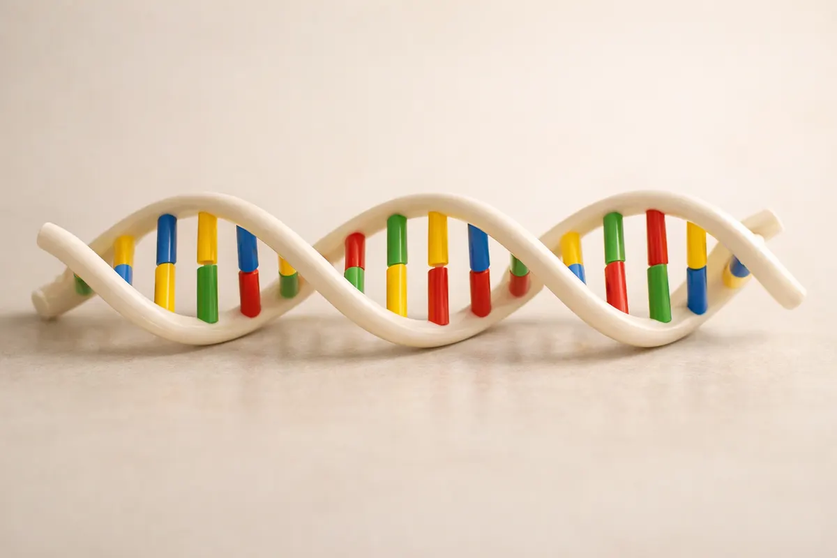
Quick answer: what base pairs are
Base pairs are the complementary relationships between nucleotide bases in double-stranded nucleic acids. In DNA, adenine (A) pairs with thymine (T) and guanine (G) pairs with cytosine (C). These pairings are held together by hydrogen bonds and the specific A–T and G–C rules determine the way two strands match to form the familiar double helix.
When people refer to "a base pair" (bp) as a unit, they frequently mean one matched A–T or G–C in double-stranded DNA. Long molecules are commonly measured in base pairs (for example, a gene that is 1,200 bp long) or in kilobases (kb) and megabases (Mb) for larger scales.
For RNA molecules that are double-stranded or fold back on themselves, uracil (U) replaces thymine, so A pairs with U instead of T. The core idea for both DNA and RNA is complementarity: each base prefers a specific partner, and that specificity is the foundation of replication, transcription, and many lab techniques.
- DNA base pairs: A–T (two hydrogen bonds), G–C (three hydrogen bonds).
- RNA pairing: A–U and G–C when double-stranded regions form.
- Base pair (bp) is both a chemical concept and a length unit for sequences.
How base pairing works and why it matters
At the chemical level, base pairing follows complementary shapes and hydrogen-bonding patterns. Adenine and thymine form two hydrogen bonds, while guanine and cytosine make three. The extra bond in G–C pairs makes those regions more thermally stable, which has practical consequences for DNA melting temperature, binding affinity, and structure.
Beyond chemistry, complementarity is a reliable encoding mechanism: the sequence on one strand determines the sequence on the other. This is the reason DNA replication copies information faithfully and why reverse-complement rules let you predict the partner strand simply by substituting bases and reversing the order.
In molecular biology, base pairs are used in many practical ways. PCR primers rely on predictable base pairing for specific binding; restriction enzymes recognize short palindromic base-pair patterns; and alignment algorithms compare base-pair sequences to identify genes, variants, and evolutionary relationships.
For students and educators, base pairs are the unit of measurement for genes and genomes. A single human chromosome can contain tens to hundreds of millions of base pairs; the haploid human genome is roughly three billion bp. For many homework and exam problems, counting bp, determining complementary strands, and calculating GC% are the core tasks.
- Complementarity enables replication, repair, and information transfer.
- G–C rich regions are more stable and melt at higher temperatures.
- The bp unit scales: bp → kb (1,000 bp) → Mb (1,000,000 bp).
Key clues
When you are given a sequence, worksheet, or a short DNA fragment, several quick checks tell you whether base pairing is consistent and whether the data make sense. Start by scanning for nonstandard letters (like N, R, Y) which indicate ambiguity; standard unambiguous DNA letters are A, T, G, and C.
Count and compare: for double-stranded DNA the number of A's on one strand equals the number of T's on the complementary strand, and G's match C's on the complementary strand. Within a single strand, however, counts can vary. Chargaff's rules (approximate equality of A≈T and G≈C in a double-stranded genome) are useful for plausibility checks on large-scale data.
Calculate GC content (percent of bases that are G or C). High GC% (>60%) suggests more stable duplexes and higher melting temperatures; low GC% (<40%) suggests lower stability. GC content also affects primer design, hybridization stringency, and interpretations of melting curves in lab assays.
Look for palindromes and repeats. Short palindromic sequences (for example GAATTC which reads CTTAAG on the complement) are recognition sites for many restriction enzymes. Repeats, homopolymers (AAAA...), and runs of ambiguous bases can cause sequencing artifacts or misaligned reads.
- Scan for invalid characters: A, T, G, C are standard; N means unknown base.
- Compute GC% = (G+C)/(A+T+G+C) × 100 to estimate duplex stability.
- Check for palindromic recognition sequences if restriction mapping is relevant.
- Compare complementary strands for A↔T and G↔C symmetry on long sequences.
Step-by-step workflow
If you need to validate a DNA sequence or solve a homework question involving base pairs, follow a concise, stepwise approach. First, confirm the input: is the sequence single-stranded letters, a reverse complement, or an electropherogram readout? Knowing the format guides the next steps.
Second, clean and normalize the sequence. Remove whitespace or line breaks, convert lowercase to uppercase, and replace ambiguous symbols (if present) with a marker. For classroom problems, treat Ns as unknowns and explain their effect on counts or calculations.
Third, compute basic metrics: total length in bp, counts of each base, GC percentage, and the reverse complement. For many questions, calculating the complementary strand and showing the base-count arithmetic is the expected solution.
Finally, interpret the results. For example, use GC% to comment on expected melting behavior, point out palindromic restriction sites if present, and flag any ambiguous or impossible patterns. When accuracy matters beyond homework—such as diagnostic or research settings—note when to escalate to experimental validation.
- Step 1: Identify the format — single strand, labeled complement, or raw read.
- Step 2: Clean sequence text and mark ambiguous bases (N, R, Y).
- Step 3: Count A/T/G/C, compute length and GC% and produce the reverse complement.
- Step 4: Report conclusions and state confidence; recommend verification for critical uses.
Practical examples of base-pair problems
Example 1 — Complement and count: Given the single-strand sequence 5'-ATGCGT-3', the reverse complement is 5'-ACGCAT-3'. The fragment length is 6 bp and counts are A:1, T:1, G:2, C:2. GC% = (2+2)/6 × 100 = 66.7%, indicating a relatively GC-rich short fragment.
Example 2 — Converting units: A gene listed as 2,500 bp equals 2.5 kb. For genome-scale measures, remember the notation: kb (kilobase) = 1,000 base pairs, Mb (megabase) = 1,000,000 base pairs. When a homework problem asks for physical length, use the average DNA rise per base pair (~0.34 nm) to convert bp to nanometers for a rough estimate, but state that this is an idealized straight-helix approximation.
Example 3 — Detecting mismatches: If a problem gives paired strands that disagree at one position (e.g., A opposite C), that is a mismatch. In biological systems mismatches can indicate sequencing error, a SNP, or damage. For classroom answers, identify the mismatch clearly and explain its implications for stability and replication fidelity.
Example 4 — RNA pairing nuance: Given an RNA hairpin region, replace T with U and check pairing A–U and G–C. Many textbook questions ask you to predict folding or stem lengths; show the paired region, count base pairs in the stem, and annotate unpaired loops.
- Compute reverse complements to check answers quickly.
- Convert between bp, kb, and Mb when reporting sizes.
- Flag mismatches as possible errors or variants, not definitive proof of biology without validation.
- For RNA, substitute U for T when checking pairing in double-stranded regions.
Limitations and when to verify base-pair conclusions
A sequence string and textbook rules let you solve many problems, but they have limits. Short fragments or ambiguous letters (N) reduce confidence in counts and complementary predictions. Sequencing technologies can introduce base-call errors, and biological samples can include mixtures of closely related sequences that a simple readout won't resolve.
Biological context matters. Knowing that a sequence comes from a mitochondrial genome, plasmid, or bacterial chromosome changes how you interpret repeats, GC content norms, and expected length. For homework, state assumptions explicitly; for real-world analyses, flag the provenance of the data and any pre-processing steps.
Thermal stability predictions from GC% are approximate. Local sequence context, salt concentration, and sequence length all influence melting temperature (Tm). For lab work such as primer design or hybridization experiments, use validated calculators or experimental validation rather than relying on GC% alone.
When the stakes are high—diagnostics, published research, or actionable clinical/forensic decisions—treat computational checks as first-pass evidence. Recommend orthogonal verification: replicate sequencing, Sanger confirmation, or consult a molecular biology lab. The app-based or homework checks described here are useful for learning and triage but not a substitute for laboratory confirmation.
- Ambiguous bases and sequencing errors reduce confidence in conclusions.
- GC% gives directional insight, not a precise melting temperature without other parameters.
- Context (organism, sample type) affects interpretation—state it when possible.
- Escalate to experimental verification for diagnostic or research-critical cases.
Related guides
Next step: check your sequence quickly with BiologyAI
If you want a fast, structured first pass on a sequence or homework problem, BiologyAI’s solver page walks you through base counts, reverse complements, GC%, and basic plausibility checks. Use it as a quick verification step after you’ve done the manual calculations above. For experiments, clinical questions, or high-stakes results, follow up with lab verification or a specialist rather than relying on an automated first pass. Visit https://biologyai.app/tools/ai-biology-solver to try the guided solver and see step-by-step explanations.


Frequently asked questions
How many base pairs are in the human genome?
The haploid human genome contains roughly 3 billion base pairs (about 3 × 10^9 bp). Because humans are diploid, most somatic cells have about 6 billion base pairs total. Reported counts vary slightly between assemblies and patches, so cite the specific genome build (for example GRCh38) if you need an exact number for a project.
What's the difference between a nucleotide and a base pair?
A nucleotide is a single subunit of DNA or RNA made of a sugar, a phosphate, and a nitrogenous base (A, T, G, C, or U). A base pair refers to two complementary bases on opposite strands that hydrogen-bond (for example A–T or G–C in DNA). When describing length, scientists often use base pairs (bp) to mean matched nucleotide pairs in double-stranded DNA.
How do I calculate GC% and why does it matter?
GC% = (number of G bases + number of C bases) ÷ (total number of A, T, G, C bases) × 100. GC% influences duplex stability because G–C pairs form three hydrogen bonds versus two for A–T pairs. Higher GC% typically raises melting temperature and affects binding stringency, primer selection, and some sequencing biases. Use GC% as an initial indicator, then apply a Tm calculator or experimental test for precise planning.
Can a photo tell me the base pairs in a sample?
A photograph of physical material (like tissue or a gel) cannot reveal base-pair sequences. Photos of gel bands can give approximate fragment sizes when compared to a ladder, but they do not provide sequence-level information. To determine specific base pairs you need sequencing data, electropherogram reads, or a written sequence. If you only have a visual cue, treat it as provisional and plan for molecular assays if sequence identity matters.