Biology AI Homework Solver: Getting Biology Help That Shows the Steps
Learn what a biology AI homework solver can and cannot infer from photos, how to check each step, and when to seek lab or teacher verification.
What a biology AI homework solver can reliably infer
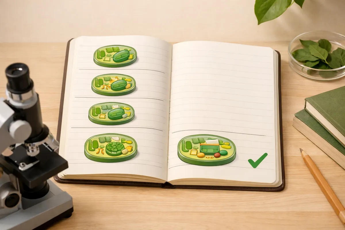
A biology AI homework solver can often walk you through a step-by-step reasoning path for visible, structural problems. For example, from a clear microscope image of a prepared slide it can describe cell shape (round vs. elongated), relative nucleus size, visible organelles like chloroplasts, and typical staining patterns. When you supply a labeled diagram, a good solver can also map diagram labels to common terms and show the steps used to reach an identification or conclusion.
Beyond direct identification, the solver excels at translating your observations into organized steps: listing the evidence (what the photo shows), matching those clues to textbook categories (e.g., plant vs. animal cell, mitosis stage based on chromosome arrangement), and showing intermediate reasoning before the final answer. That stepwise trace — evidence, comparisons, rule application, and tentative conclusion — is exactly what makes an AI solver useful for homework rather than a black-box answer generator.
For calculation-style biology problems (population growth curves, Hardy-Weinberg practice, simple dilution math), an AI can show the formulas, substitute numbers from your problem statement or image, and present each arithmetic step. If the question is about experimental design or predicting outcomes from a described setup, the solver can list assumptions, expected controls, and a logical prediction chain based on common biological principles.
Use the app as a method-checker: ask it to show its steps, then compare those steps to your notes or textbook. When the solver flags alternative interpretations (for example, two species that look similar in the same photo), that multiple-hypothesis output is valuable because it tells you which clues to re-check rather than presenting one confident but unsupported label.
- Visible morphology: cell shape, tissue arrangement, gross organ structure from clear photos.
- Staining and contrast patterns: nuclei, cell walls, and chloroplasts when staining or lighting reveals them.
- Stage-based identification: mitosis phases or visible lifecycle stages when chromosomes or structures are clear.
- Stepwise calculations: show formulas and arithmetic for genetics, ecology, and growth problems.
- Hypothesis lists: multiple plausible IDs with the deciding clues noted rather than a single final claim.
What a biology AI homework solver cannot prove from photos
A biology AI homework solver cannot conclusively prove chemical purity, pathogenicity, toxicity, or species identity when those require lab tests, DNA sequencing, or culture. For example, two bacterial species with similar colony morphology can look nearly identical in photos but behave differently under biochemical tests and staining protocols. Any claim that suggests absolute certainty about safety, toxicity, or authenticity from a single photo is outside the solver’s scope.
Color alone is an unreliable indicator. Camera white balance, lighting, and specimen preservation all change perceived color. Many lookalikes in biology differ by subtle anatomical measurements, microscopic features, or biochemical markers that photographs—especially casual phone photos—do not capture. The solver can point out plausible lookalikes, but it cannot replace assays, microscope slides at required magnifications, or professional lab diagnostics.
A solver also cannot verify that a student’s experimental procedure was actually followed or that data were collected without bias. It can assess described methods and highlight likely sources of error, but it has no access to raw lab conditions, reagent quality, or procedural nuances that influence experimental validity. Treat its critique as guidance to check, not as certification of lab rigor.
Finally, the AI cannot take the place of instructor judgment when academic integrity or grading policies are involved. Use the step-by-step explanations to learn and validate your reasoning, but do not submit AI-generated conclusions as your own work without adapting and confirming them against course requirements and instructor feedback.
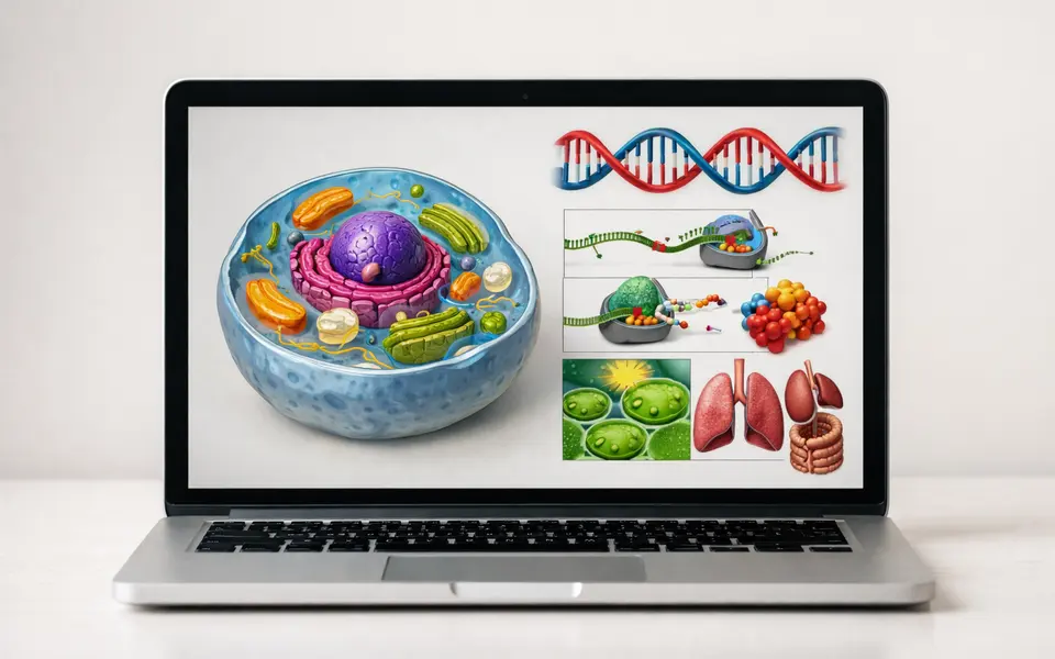
- Cannot confirm species identity that requires DNA, biochemical tests, or microscopy beyond the photo’s resolution.
- Cannot prove safety, toxicity, or pathogenicity from images alone.
- Cannot certify experimental procedure, reagent integrity, or lab-grade measurements.
- Cannot replace instructor review or official grading; use outputs as study support.
Practical visual clues to capture before using a solver
Good answers start with good evidence. Before you run a photo through a biology AI homework solver, gather images that emphasize stable, reproducible clues: overall shape and scale, context (is the specimen in tissue, whole organism, or slide), and at least one scale reference such as a ruler, a stage micrometer, or a coin. A single close-up and a single contextual shot together often reveal more than multiple similar close-ups.
Lighting and focus matter more than saturation. Natural diffuse light or ring lights reduce harsh shadows that hide structural details; bright built-in flash often blows out highlights and erases texture. Capture the same subject from multiple angles if asymmetry matters (for example, leaf underside vs. top surface) and include a plain background to avoid confusing pattern recognition.
Microscope photos require scale and staining info. If you’re photographing a slide, include the magnification (10x, 40x) and the stain used (methylene blue, iodine). Those two data points let a solver map visible features to the right interpretation: what looks like a large organelle at 10x might be something else at 40x. For diagrams or textbook images, include the problem text or labels as a separate photo so the solver can tie image evidence to the question prompt.
Finally, capture metadata and context: where the specimen was found (pond edge, forest floor, classroom culture), approximate size, and behavior if applicable (movement pattern, response to stimulus). These non-visual details often shift the solver’s ranked hypotheses more than tiny color variations.
- Include scale: ruler, coin, or known magnification in microscope images.
- Shoot one contextual and one close-up image to show both environment and detail.
- Note magnification and stains on slide photos to avoid misinterpretation.
- Use diffuse lighting and a plain background to preserve texture and structure.
- Add short contextual notes: location, size estimate, and observed behavior.
When to verify AI steps with tests, teachers, or labs
Treat the solver’s output as a prioritized checklist for verification. If the AI lists multiple plausible IDs, use that list to decide what to test next. For species-level uncertainty in classroom assignments, ask your instructor which verification they accept: a higher-magnification image, a staining procedure, or a short culture and biochemical test. Many teachers accept a clear explanation of why you favored one hypothesis and what follow-ups you would run to confirm it.
Use simple in-class tests before sending anything to a lab. For example, iodine can help distinguish starch-containing plant cells, and a wet mount can reveal movement in protozoa. These are low-cost checks that strengthen or refute the solver’s steps. When chemical or pathogenic safety is a concern, stop and seek professional lab testing rather than relying on photos or home kits that are not validated for diagnostic use.
When health, safety, or legal concerns arise (possible pathogens, toxic fungi, regulated organisms), contact a qualified lab, public health authority, or your teacher immediately. A biology AI homework solver’s role ends where biosafety, clinical diagnosis, or legal regulation begins. The app can prepare a clear evidence packet—images, solver steps, and suggested confirmatory tests—to hand to professionals, which speeds official verification.
For grading and integrity questions, use the solver to show your working: include the solver’s step list as a study aid, then rewrite the steps in your own words and cite textbook or lecture sources where appropriate. If you’re unsure which parts of the AI’s reasoning are acceptable in your course, check with your instructor before submission. That preserves both learning and academic honesty.
- Ask your teacher which confirmations they accept (higher magnification, staining, or a short assay).
- Run low-risk in-class checks (wet mounts, iodine tests) to strengthen visual IDs.
- Escalate to certified labs for pathogenicity, toxicity, DNA-based species ID, or regulated organisms.
- For safety concerns, contact public health or a qualified lab rather than relying on photo analysis.
- Use AI steps as study aid: rewrite them in your own words and cite sources before submitting.
Related guides
Use BiologyAI to check your steps, not to skip them
Try BiologyAI as a first-pass study partner: take clear context and close-up photos, ask the solver to show each reasoning step, and use that step list to test or photograph the decisive clues the app highlights. If a claim touches safety, species-level ID, or diagnostic certainty, follow the solver’s verification path—ask your teacher, run an in-class check, or seek lab confirmation. Download the BiologyAI app to see stepwise explanations on iOS and to turn your photos and notes into a systematic checklist you can verify before submitting or acting on any conclusion.


Frequently asked questions
Can a biology AI homework solver grade my lab report for me?
An AI solver can help you check methods, point out likely sources of error, and suggest clearer ways to present steps and calculations. It should not be used to produce or submit work as your own. Treat its feedback as a study tool: apply suggested corrections, rewrite explanations in your own voice, and confirm any substantive changes with your instructor before turning in graded work.
How accurate are species IDs from phone photos?
Accuracy varies widely: clear photos showing diagnostic features and scale can support reliable IDs at genus or family level in many cases, but species-level confirmation often requires microscopic detail, measurements, or genetic tests. If the solver gives multiple plausible species, follow up with higher-resolution imaging, specific staining, or a lab test when species-level accuracy matters.
Is it safe to rely on the solver to decide if a specimen is poisonous or harmful?
No. The solver can flag lookalikes and list cautious warnings, but it cannot certify toxicity or safety from images alone. When safety is in question, stop handling the specimen, consult your teacher or local experts, and use professional testing or established field guides. For potentially harmful organisms, prioritize expert verification over photo-based conclusions.
What should I include in my photos to get the most useful step-by-step answer?
Include a scale reference, a contextual shot showing the specimen’s environment, a close-up of diagnostic features, and any relevant labels or the problem text. Note magnification and stains for microscope images, and add short context like size estimate and where it was found. These details let the solver show clear intermediate steps tied to specific evidence.